Home
/ Abdomen Anatomy Female : Female Lower Abdominal Organs Download Scientific Diagram - Anatomy of the female abdomen and pelvis medical illustration.
Abdomen Anatomy Female : Female Lower Abdominal Organs Download Scientific Diagram - Anatomy of the female abdomen and pelvis medical illustration.
Abdomen Anatomy Female : Female Lower Abdominal Organs Download Scientific Diagram - Anatomy of the female abdomen and pelvis medical illustration.. Anatomy, abdomen and pelvis, female internal genitals; Female abdominal anatomy images female abdominal anatomy. The abdomen (commonly called the belly) is the body space between the thorax (chest) and pelvis. Among all the pain in lower right abdominal anatomy, the female is a very familiar topic. The major organs of the abdomen include the small intestine, large intestine, and stomach.
Is a health blogger focusing on health, beauty, lifestyle and fitness topics. The right ovary and fallopian tube are also located in the right lower quadrant in females. In women, the lowest portion of the abdomen is actually the pelvis and involves the uterus,. Take free questions on this article. The abdomen (commonly called the belly) is the body space between the thorax (chest) and pelvis.
Diagram Of The Abdomen Koibana Info Anatomie Des Organes Anatomie Corps Humain Anatomie Du Corps from i.pinimg.com This landmark begins at the level of the sacral promontory posteriorly and the pubic symphysis anteriorly. The major organs of the abdomen include the small intestine, large intestine, and stomach. Take free questions on this article. Anatomy of neck vein 12 photos of the anatomy of neck vein anatomy internal jugular vein cannulation, anatomy of internal jugular vein in neck, anatomy of internal jugular vein pdf, anatomy of jugular vein in cattle, anatomy of the neck veins, human anatomy, anatomy internal jugular vein cannulation, anatomy of internal. The abdomen (colloquially called the belly, tummy, midriff or stomach) is the part of the body between the thorax (chest) and pelvis, in humans and in other vertebrates. At the level of the pelvic bones, the abdomen. The right lower quadrant contains the appendix and cecum in both males and females. We're going to take apart a plastic anatomy model and see what we can find in the abdomen.
Affordable and search from millions of royalty free images, photos and vectors.
We're going to take apart a plastic anatomy model and see what we can find in the abdomen. At the level of the pelvic bones, the abdomen. This landmark begins at the level of the sacral promontory posteriorly and the pubic symphysis anteriorly. Among all the pain in lower right abdominal anatomy, the female is a very familiar topic. The abdominal cavity is the part of the body that houses the stomach, liver, pancreas, kidneys, gallbladder, spleen, and the large and small intestines. The major organs of the abdomen include the small intestine, large intestine, and stomach. The diaphragm forms the upper surface of the abdomen. Anatomy of the female abdomen and pelvis medical illustration. The liver, stomach, and abdominal contents are clearly identified and labeled, including the cecum, ascending colon, transverse colon, descending colon, and small intestine. The abdomen (colloquially called the belly, tummy, midriff or stomach) is the part of the body between the thorax (chest) and pelvis, in humans and in other vertebrates. The diaphragm forms the upper surface of the abdomen. Gallbladder pain is below the ribs on the upper right side of the abdomen. Abdominal anatomy chart female, abdominal anatomy female diagram, anatomy of female abdomen and pelvis, anatomy of female abdominal cavity, anatomy of female human.
Take free questions on this article. Stomach is a muscular bag forming the most distensible part of the human digestive system. Anatomy, abdomen and pelvis, female external genitalia. 1 the bladder is supported by ligaments and connects at the top to two ureters. Together, these three turn nutrients into usable energy, as well as help dispose of solid waste.
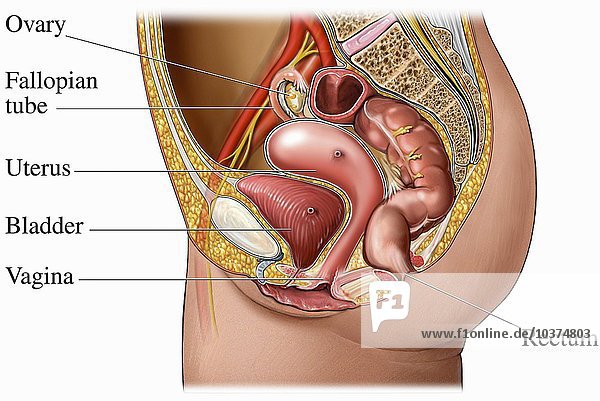
Medical Illustration Of The Normal Anatomy Of The Human Female Abdomen And Pelvis From A Lateral Cut Away View from www1.f1online.de The liver, stomach, and abdominal contents are clearly identified and labeled, including the cecum, ascending colon, transverse colon, descending colon, and small intestine. Among all the pain in lower right abdominal anatomy, the female is a very familiar topic. The abdomen (commonly called the belly) is the body space between the thorax (chest) and pelvis. Together, these three turn nutrients into usable energy, as well as help dispose of solid waste. The kidneys, the ureters, the bladder, and the urethra (fig. The space below contains the bladder, rectum, and part of the descending colon. Abdominal computed tomography (ct) is a type of medical imaging procedure used to diagnose and monitor internal stomach issues, like cancer, bowel obstruction, and abdominal pain. At the level of the pelvic bones, the abdomen.
Anatomy, abdomen and pelvis, female internal genitals;
Abdominal anatomy chart female, abdominal anatomy female diagram, anatomy of female abdomen and pelvis, anatomy of female abdominal cavity, anatomy of female human. Find the perfect abdominal anatomy stock photos and editorial news pictures from getty images. The major organs of the abdomen include the small intestine, large intestine, and stomach. The right ovary and fallopian tube are also located in the right lower quadrant in females. At the level of the pelvic bones, the abdomen. In women the lowest portion of the abdomen is actually the pelvis and involves the uterus fallopian tubes and ovaries. Anatomy, abdomen and pelvis, female external genitalia. The space below contains the bladder, rectum, and part of the descending colon. Related posts of anatomy of the abdomen women anatomy of neck vein. The liver, stomach, and abdominal contents are clearly identified and labeled, including the cecum, ascending colon, transverse colon, descending colon, and small intestine. The female urinary tract is comprised of four organs: The major muscles of the abdomen include the rectus abdominis in front the external obliques at the sides and the latissimus dorsi muscles in the back. Those organs include the stomach, small intestine, colon, liver, gallbladder, spleen, and pancreas.
Child physical examination concept female pediatrician or health care practitioner examines baby girl's abdomen. The major muscles of the abdomen include the rectus abdominis in front the external obliques at the sides and the latissimus dorsi muscles in the back. The abdomen (colloquially called the belly, tummy, midriff or stomach) is the part of the body between the thorax (chest) and pelvis, in humans and in other vertebrates. Female reproductive anatomy focuses on two groups of organs; The true pelvis, or lesser pelvis, lies below the pelvic brim (figure 1).
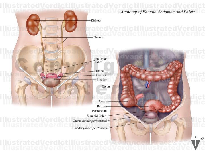
Stock Female Pelvis Normal Anatomy Illustrated Verdict from images.squarespace-cdn.com The diaphragm forms the upper surface of the abdomen. Among all the pain in lower right abdominal anatomy, the female is a very familiar topic. The female urinary tract is comprised of four organs: Anatomy of the female abdomen and pelvis medical illustration. This landmark begins at the level of the sacral promontory posteriorly and the pubic symphysis anteriorly. In women the lowest portion of the abdomen is actually the pelvis and involves the uterus fallopian tubes and ovaries. The regions occupied by stomach are epigastric, umbilical and hypochondriac regions. The muscles of the lower back, including the erector spinae and quadratus lumborum muscles, contract to extend and laterally bend the vertebral column.
The major organs of the abdomen include the small intestine, large intestine, and stomach.
In women, the bladder is bordered posteriorly by the uterus and vagina. Anatomy of neck vein 12 photos of the anatomy of neck vein anatomy internal jugular vein cannulation, anatomy of internal jugular vein in neck, anatomy of internal jugular vein pdf, anatomy of jugular vein in cattle, anatomy of the neck veins, human anatomy, anatomy internal jugular vein cannulation, anatomy of internal. The kidneys, the ureters, the bladder, and the urethra (fig. We're going to take apart a plastic anatomy model and see what we can find in the abdomen. The right ovary and fallopian tube are also located in the right lower quadrant in females. The abdomen (commonly called the belly) is the body space between the thorax (chest) and pelvis. Find the perfect abdominal anatomy stock photos and editorial news pictures from getty images. Anatomy, abdomen and pelvis, female internal genitals; Stomach is a muscular bag forming the most distensible part of the human digestive system. 1 the bladder is supported by ligaments and connects at the top to two ureters. The right lower quadrant contains the appendix and cecum in both males and females. He has been with healthiack.com since 2012 and has written and reviewed well over 500 coherent articles. Child physical examination concept female pediatrician or health care practitioner examines baby girl's abdomen.
Related posts of anatomy of the abdomen women female bone structure ideas abdomen anatomy-female. Is a health blogger focusing on health, beauty, lifestyle and fitness topics.
